February 2, 2012

Rituximab possible treatment option for patients with primary biliary cirrhosis

Public release date: 2-Feb-2012

Contact: Dawn Peters
healthnews@wiley.com
781-388-8408
Wiley-Blackwell

Drug found to be safe in PBC patients not responsive to ursodeoxycholic acid

An open-label study of rituximab, a monoclonal antibody for human CD20, was shown to be safe in patients with primary biliary cirrhosis (PBC) who had an incomplete response to the standard ursodeoxycholic acid (UDCA) therapy, also known as Ursodiol. Study details available in the February issue of Hepatology, a journal published by Wiley-Blackwell on behalf of the American Association for the Study of Liver Diseases, report that rituximab was successful in reducing the level of alkaline phosphatase (ALP)—a protein used to measure liver injury.

According to the National Institute of Diabetes and Digestive and Kidney Diseases (NIDDK), PBC—an autoimmune liver disease characterized by inflammation of the bile ducts that ultimately causes bile to build up and damage the liver—typically occurs between the ages of 40 and 60, primarily affecting women. Medical studies have identified the presence of anti-mitochondrial autoantibodies (AMA) to enzymes involved in the production of the body's energy (pyruvate dehydrogenase complex-PDC-E2) in up to 95% of PBC cases.

Currently, the standard therapy for PBC is UDCA or liver transplantation in patients who have progressed to end-stage liver disease. However, previous studies have shown that UDCA may be ineffective in up to 40% of PBC patients and 10% could require transplantation or die from the disease. "Small trials using immunosuppressants have failed to demonstrate significant clinical benefit or carry unacceptable safety profiles," said Dr. Christopher Bowlus with UC Davis Health System in California and lead author of the current study.

Dr. Bowlus added, "Given our previous work implicating B cells to the development of this disease, we hypothesized that a therapy such as rituximab, which depletes B cells, could offer a potentially effective treatment option with acceptable adverse effects." The team enrolled 6 patients between the ages of 18 and 65 years of age who were diagnosed with PBC and had an incomplete response to UDCA despite 6 months of therapy. Participants in this open-label study received an intravenous infusion of 1000 mg of rituximab on day 1 and day 15, with follow-up for 52 weeks. Two patients received one dose of rituximab due to latent varicella (chickenpox) activation in one and viral respiratory infection in the other.

The trial results found that rituximab was safe and well tolerated by PBC patients, with no serious adverse events reported. By 16 weeks post-treatment patients had significantly lower levels of immunoglobulins IgG, IgM, and IgA, which are the antibodies normally present in blood, but in the case of IgM are often elevated in PBC. In addition, the abnormal antibodies directed against mitochondria (AMA), were also reduced after treatment. However, levels returned to baseline by 36 weeks. Further, serum alkaline phosphatase decreased up to 36 weeks post-treatment.

The authors noted that rituximab therapy could clinically improve PBC through multiple pathways that include the reduction of anti-mitochondrial antibodies through depletion of memory B cells, increases in regulatory T cells associated with immune response, and modulation of cytokine production involved with inflammation. "Further investigation of B cell targeting strategies is necessary to develop potentially novel therapeutic options for patients with PBC," concludes Dr. Bowlus.

###

Full Citation: Biochemical and Immunologic Effects of Rituximab in Primary Biliary Cirrhosis Patients with an Incomplete Response to Ursodeoxycholic Acid." Masanobu Tsuda, Yuki Moritoki, Zhe-Xiong Lian, Weici Zhang, Katsunori Yoshida, Kanji Wakabayashi, Guo-Xiang Yang, Toshio Nakatani, John Vierling, Keith Lindor, M. Eric Gershwin and Christopher L. Bowlus. Hepatology; Published Online: January 27, 2012 (DOI: 10.1002/hep.24748); Print Issue Date: February 2012. http://onlinelibrary.wiley.com/doi/10.1002/hep.24748/abstract.

Author Contact: To arrange and interview with Dr. Bowlus, please contact Charles Casey with the Office of Public Affairs at UC Davis Health System at charles.casey@ucdmc.ucdavis.edu or 916-734-9048.

This study is published in Hepatology. Media wishing to receive a PDF of the article may contact healthnews@wiley.com.

About the Journal

Hepatology is the premier publication in the field of liver disease, publishing original, peer-reviewed articles concerning all aspects of liver structure, function and disease. Hepatology's current impact factor is 10.885.Each month, the distinguished Editorial Board monitors and selects only the best articles on subjects such as immunology, chronic hepatitis, viral hepatitis, cirrhosis, genetic and metabolic liver diseases and their complications, liver cancer, and drug metabolism. Hepatology is published on behalf of the American Association for the Study of Liver Diseases (AASLD). For more information, please visit http://onlinelibrary.wiley.com/journal/10.1002/(ISSN)1527-3350 .

About Wiley-Blackwell

Wiley-Blackwell is the international scientific, technical, medical, and scholarly publishing business of John Wiley & Sons, with strengths in every major academic and professional field and partnerships with many of the world's leading societies. Wiley-Blackwell publishes nearly 1,500 peer-reviewed journals and 1,500+ new books annually in print and online, as well as databases, major reference works and laboratory protocols. For more information, please visit http://www.wileyblackwell.com or our new online platform, Wiley Online Library (http://www.wileyonlinelibrary.com), one of the world's most extensive multidisciplinary collections of online resources, covering life, health, social and physical sciences, and humanities.

Source

Coffee consumption reduces fibrosis risk in those with fatty liver disease

medxpress-logo

February 2, 2012

Caffeine consumption has long been associated with decreased risk of liver disease and reduced fibrosis in patients with chronic liver disease. Now, newly published research confirms that coffee caffeine consumption reduces the risk of advanced fibrosis in those with nonalcoholic fatty liver disease (NAFLD). Findings published in the February issue of Hepatology, a journal of the American Association for the Study of Liver Diseases, show that increased coffee intake, specifically among patients with nonalcoholic steatohepatitis (NASH), decreases risk of hepatic fibrosis.

The steady increase in rates of diabetes, obesity, and metabolic syndrome over the past 20 years has given rise to greater prevalence of NAFLD. In fact, experts now believe NAFLD is the leading cause of chronic liver disease in the U.S., surpassing both hepatitis B and C. The majority of patients will have isolated fatty liver which has a very low likelihood of developing progressive liver disease. However, a subset of patients will have NASH, which is characterized by inflammation of the liver, destruction of liver cells, and possibly scarring of the liver. Progression to cirrhosis (advanced scarring of the liver) may occur in about 10-11% of NASH patients over a 15 year period, although this is highly variable.

To enhance understanding of the correlation between coffee consumption and the prevalence and severity of NAFLD, a team led by Dr. Stephen Harrison, Lieutenant Colonel, U.S. Army at Brooke Army Medical Center in Fort Sam Houston, Texas surveyed participants from a previous NAFLD study as well as NASH patients treated at the center's hepatology clinic. The 306 participants were asked about caffeine coffee consumption and categorized into four groups: patients with no sign of fibrosis on ultrasound (control), steatosis, NASH stage 0-1, and NASH stage 2-4.

Researchers found that the average milligrams in total caffeine consumption per day in the control, steatosis, Nash 0-1, and Nash 2-4 groups was 307, 229, 351 and 252; average milligrams of coffee intake per day was 228, 160, 255, and 152, respectively. There was a significant difference in caffeine consumption between patients in the steatosis group compared to those with NASH stage 0-1. Coffee consumption was significantly greater for patients with NASH stage 0-1, with 58% of caffeine intake from regular coffee, than with NASH stage 2-4 patients at only 36% of caffeine consumption from regular coffee.

Multiple analyses showed a negative correlation between coffee consumption and risk of hepatic fibrosis. "Our study is the first to demonstrate a histopatholgic relationship between fatty liver disease and estimated coffee intake," concludes Dr. Harrison. "Patients with NASH may benefit from moderate coffee consumption that decreases risk of advanced fibrosis. Further prospective research should examine the amount of coffee intake on clinical outcomes."

More information: "Association of Coffee and Caffeine Consumption with Fatty Liver Disease, Non-alcoholic Steatohepatitis, and Degree of Hepatic Fibrosis." Jeffrey W Molloy, Christopher J Calcagno, Christopher D Williams, Frances J Jones, Dawn M Torres, Stephen A Harrison. Hepatology; December 22, 2011 (DOI: 10.1002/hep.24731); Print Issue Date: February 2012.

Provided by Wiley (news : web)

Source

February 1, 2012

Straight from the gut: Microbes can cause obesity

stomach-vertical

01/02/2012 18:20:00

By Helen Dodson - Obesity and chronic liver disease can be triggered by a family of proteins that alter populations of microbes in the stomach, a discovery that suggests the condition may be infectious,

Yale scientists report. The study, in the advance online publication of Nature, expands on earlier Yale research that showed how similar microbial imbalances caused by the same family of proteins increases the risk of intestinal diseases such as colitis.

The Yale scientists’ most extraordinary finding, they said, was that the altered intestinal environment that led to obesity and liver disease was infectious among the community of mice. “When healthy mice were co-housed with mice that had altered gut microbes, the healthy mice also developed a susceptibility for development of liver disease and obesity,” said senior author Richard A. Flavell, professor of immunobiology at Yale School of Medicine and a Howard Hughes Medical Institute investigator.

The proteins in question are called inflammasomes. They are responsible for launching the immune system’s inflammatory response. Inflammasomes act as sensors and regulators of the microbial environment of the intestines.

The Yale team found that a deficiency in components of two particular inflammasomes in mice resulted in the development of an altered microbial community associated with increased bacteria. This determined the severity of non-alcoholic fatty liver disease (NAFLD) and obesity in the mice.

NAFLD is the result of metabolic syndrome, a collection of disorders that includes obesity and diabetes, and is the leading cause of chronic liver disease in the western world. It is estimated that up to 30 million people suffer from NAFLD in the United States alone. Twenty percent of people with NAFLD develop chronic liver inflammation, placing them at risk for cirrhosis and liver cancer, but the causes have been unclear.

The next step, Flavell said, is extending this research to humans and to identify more precisely the bacteria involved in the progression to liver disease. “We found, in mice, that targeted antibiotic treatment brought the microbial composition back to normal, and thus eased the liver disease. Our hope is that our findings may eventually lead to a treatment for humans.”

The researchers who led this project in Flavell’s laboratory are Jorge Henao-Mejia, Eran Elinav, and Chengcheng Jin. Other participating researchers were Liming Hao, Wajahat Z. Mehal, Till Strowig, Christoph A. Thaiss, Stephanie C. Eisenbarth, Michael J. Jurczak, Joao-Paulo Camporez, and Gerald I. Shulman of Yale; Andrew L. Kau and Jeffrey I. Gordon of Washington University School of Medicine; and Hal M. Hoffman of the University of California at San Diego.

The study was supported by grants from the Howard Hughes Medical Institute, United States-Israel Binational Foundation, the Crohn’s and Colitis Foundation of America, the National Institutes of Health, a VA Merit Award, and the Claire and Emmanuel G. Rosenblatt Award from the American Physicians for Medicine in Israel Foundation. Postdoctoral fellowships were provided by The Cancer Research Institute, and the Leukemia and Lymphoma Society.

For more information on Flavell’s research, visit his website.

Contact Helen Dodson helen.dodson@yale.edu 203-436-3984

Source

Current Recommendations for Using Telaprevir and Boceprevir in Patients With Advanced Fibrosis or Cirrhosis

Expert Commentary

Paul J. Pockros, MD

Posting Date: December 19, 2011

Head, Division of Gastroenterology/Hepatology
Director, SC Liver Research Consortium
Clinical Director of Research, Scripps Translational Science Institute
The Scripps Clinic
La Jolla, California

Editor’s note: In this edition of Journal Options Hepatitis, we feature the 5 pivotal phase III studies that led to the approval in 2011 of boceprevir and telaprevir for the treatment of chronic hepatitis C. Each commentary in this series addresses a key issue or question of clinical relevance related to the use of these agents in clinical practice.

Patients with cirrhosis or advanced fibrosis due to hepatitis C virus (HCV) are a particularly challenging group to treat with combination therapy that includes 1 of the currently approved direct-acting antivirals (DAA), boceprevir or telaprevir. Patients with decompensated cirrhosis have the greatest need for curative therapy; however, these individuals were not studied in the pivotal trials of boceprevir and telaprevir and are not included in the prescribing information for either drug; therefore, there is significant risk associated with treating this group of patients in the absence of experience or guidelines.[1-3] Although patients with compensated cirrhosis were included in the phase III trials of both telaprevir and boceprevir, the number of these patients is too small on which to base treatment decisions with confidence. Furthermore, patients with cirrhosis who failed previous therapy—individuals comprising a significant proportion of our current patient population—do not respond as well as others to triple therapy and will often develop protease inhibitor–resistant variants at the time of treatment failure.[4]

So how should clinicians go about implementing telaprevir and boceprevir as treatment in patients with advanced fibrosis or cirrhosis? Here I describe what we know about telaprevir and boceprevir in patients with advanced liver disease based on data from the pivotal clinical trials, along with how my colleagues and I currently go about treating these individuals in clinical practice.

Compensated Cirrhosis
Implementing Telaprevir and Boceprevir
The combined data for patients with compensated cirrhosis in all 3 phase III trials of telaprevir revealed an overall sustained virologic response (SVR) rate of 62%, and combined data in fixed-duration and response-guided arms for boceprevir demonstrated an SVR rate of 48%, rates which are certainly high enough to warrant treating compensated cirrhosis.[5-9] Notably, the addition of IL28B testing does not provide sufficient specificity to aid in predicting which cases are likely to fail treatment, and thus we do not use this routinely in my practice for cirrhotic patients.[10,11]

Data from the REALIZE trial showed much lower SVR rates with telaprevir-based therapy among previous null responders to peginterferon/ribavirin with cirrhosis (14%) or bridging fibrosis (30%).[7] Similar data are not available for boceprevir because of the exclusion of null responders in the RESPOND-2 trial.[9] Subanalysis of the arm from REALIZE that received 4 weeks of lead-in treatment with peginterferon/ribavirin before addition of telaprevir indicated that a < 1 log10 IU/mL decrease in HCV RNA at Week 4 was associated with treatment failure in patients with compensated cirrhosis, whereas a ≥ 1 log10 IU/mL decrease in HCV RNA was associated with SVR in approximately 50%.[12] Although it is not recommended in the prescribing information for telaprevir, based on these findings, my colleagues and I routinely employ a 4-week peginterferon/ribavirin lead-in for all null responder patients with advanced liver fibrosis, and we do not initiate telaprevir until the HCV RNA value at Week 4 has been reviewed. For treatment-experienced patients lacking interferon sensitivity, we defer therapy for future clinical trials of quadruple therapy or interferon-free regimens (eg, daclatasvir, asunaprevir, and peginterferon/ribavirin; PSI-7977 plus ribavirin; others).[13]

Similarly, when planning to use boceprevir in patients with compensated cirrhosis, my colleagues and I implement the 4-week peginterferon/ribavirin lead-in phase, as indicated in the boceprevir prescribing information.[2] We wait to see the HCV RNA results at Week 4 before deciding whether to expose patients to boceprevir. If patients do not have at least a 1-log10 reduction in HCV RNA from baseline, we defer therapy or enroll patients in clinical trials. Other experts follow the recommendations in the prescribing information and continue therapy until the 12-week futility rule evaluation point and use response at this time point to determine whether treatment should be continued.

Duration of Therapy
Although we have no published data regarding the benefit of extending peginterferon/ribavirin therapy to 48 weeks in cirrhotic patients who achieve an extended rapid virologic response (ie, undetectable HCV RNA at Weeks 4 and 12) on telaprevir/peginterferon/ribavirin, the telaprevir prescribing information provides a small amount of data on this issue. Of 30 patients with cirrhosis who achieved an extended rapid virologic response, 67% (12 of 18) attained SVR when the duration of peginterferon/ribavirin was shortened to 24 weeks, and 92% (11 of 12) attained SVR when peginterferon/ribavirin was administered for the full 48 weeks.[1] These are very small numbers on which to base treatment decisions, and we need more robust studies in cirrhotics to evaluate the duration of peginterferon/ribavirin therapy when combined with telaprevir. Because these data are not yet available, I administer peginterferon/ribavirin for the full 48 weeks in cirrhotic patients if they can tolerate it; I shorten therapy to 24 weeks if they cannot.

With regard to boceprevir, the prescribing information clearly indicates that patients with compensated cirrhosis should receive 4 weeks of peginterferon/ribavirin followed by 44 weeks of boceprevir in combination with peginterferon/ribavirin.[2] This is based on data from clinical trials that clearly show a benefit of fixed-duration rather than response-guided therapy in this population.[2] Although the numbers are again small, among treatment-naive cirrhotic patients, SVR rates were 42% (10 of 24) with a fixed-duration 48-week regimen vs 31% (5 of 16) when response-guided therapy was employed. Among treatment-naive individuals, SVR rates were 77% (17 of 22) and 35% (6 of 17), respectively, with fixed vs response-guided therapy. Thus, when treating with boceprevir, I administer the recommended 48 weeks of peginterferon/ribavirin in cirrhotic patients if they can tolerate it. If the patient is unable to tolerate 48 weeks of peginterferon plus ribavirin, we push duration as long as possible to that point, but at least 24 weeks. It is expected that shortened durations of therapy would compromise efficacy.

Dosing
No dosage adjustment of boceprevir is recommended for patients with mild, moderate, or severe hepatic impairment.[2] Dose modification of telaprevir is not required when it is administered to patients with mild hepatic impairment (Child-Pugh A, score 5-6), although a 15% reduction in steady-state exposure was observed in HCV-negative subjects with mild hepatic impairment compared with healthy subjects.[1] When my colleagues and I treat patients with compensated cirrhosis with telaprevir, we do not adjust the telaprevir dosage. However, when treating patients with compensated cirrhosis with either protease inhibitor, we monitor weekly for expected reductions in white and red blood cell counts, and we implement higher thresholds for reducing the dosage of peginterferon or ribavirin when declines in absolute neutrophil count and hemoglobin occur. Specifically, we will normally dose-reduce ribavirin for hemoglobin levels < 10 g/dL and peginterferon for an absolute neutrophil count < 500.For more information on anemia management, see the accompanying commentary by Brian Pearlman.

Decompensated Cirrhosis
Telaprevir is not recommended for use in patients with moderate or severe hepatic impairment (Child-Pugh B or C, score ≥ 7).[1] My colleagues and I have selected a few patients with a prior history of a single decompensation event (eg, a remote history of variceal bleeding followed by stability and low Model of End-Stage Liver Disease scores for years) to undergo triple therapy with telaprevir/peginterferon/ribavirin. This was done only after patients completed a transplant evaluation and were approved and/or listed. Thus far, 3 of 6 patients have decompensated (1 from hepatic encephalopathy, 2 because of new-onset ascites with spontaneous bacterial peritonitis), likely due to the peginterferon component of the regimen. All 3 patients were hospitalized and treatment was stopped; all recovered.

The safety and efficacy of boceprevir have not been studied in patients with decompensated cirrhosis, and the poor safety and tolerability of peginterferon/ribavirin in patients with decompensated cirrhosis remains a contraindication to treatment in this population.[8,9] My colleagues and I have not yet treated patients with decompensated cirrhosis with boceprevir. However, the data in cirrhotics in the pivotal trials of boceprevir were equally as good as those with telaprevir, so we are currently beginning to start patients on this regimen.

To date, treatment with protease inhibitor–based therapy in decompensated cirrhotics cannot be recommended outside of centers highly experienced in the management of this patient population.

Please review the remaining 4 commentaries in this series on the use of boceprevir and telaprevir in clinical practice:

  • To review strategies for management of telaprevir-associated rash and anorectal symptoms, click here.
  • For a better understanding of futility rules and their importance with boceprevir and telaprevir, click here.
  • To review the impact of the occurrence and management of anemia with boceprevir and telaprevir, click here.
  • To review rules for following response-guided therapy guidelines with telaprevir and boceprevir, click here.
References

1. Incivek [package insert]. Cambridge, Mass: Vertex Pharmaceuticals Inc.; 2011.

2. Victrelis [package insert]. Whitehouse Station, NJ: Merck & Co, Inc.; 2011.

3. Ghany MG, Nelson DR, Strader DB, et al. An update on treatment of genotype 1 chronic hepatitis C virus infection: 2011 practice guideline by the American Association for the Study of Liver Diseases. Hepatology. 2011;54:1433-1444.

4. Pockros PJ. Drugs in development for viral hepatitis: care and caution. Drugs. 2011;71:263-271.

5. Jacobson IM, McHutchison JG, Dusheiko G, et al. Telaprevir for previously untreated chronic hepatitis C virus infection. N Engl J Med. 2011;364:2405-2416.

6. Sherman KE, Flamm SL, Afdhal NH, et al. Response-guided telaprevir combination treatment for hepatitis C virus infection. N Engl J Med. 2011;365:1014-1024.

7. Zeuzem S, Andreone P, Pol S, et al. Telaprevir for retreatment of HCV infection. N Engl J Med. 2011;364:2417-2428.

8. Poordad F, McCone J, Bacon BR, et al. Boceprevir for untreated chronic HCV genotype 1 infection. N Engl J Med. 2011;364:1195-1206.

9. Bacon BR, Gordon SC, Lawitz E, et al. Boceprevir for previously treated chronic HCV genotype 1 infection. N Engl J Med. 2011;364:1207-1217.

10. Pol S, Aerssens J, Zeuzem S, et al. Similar SVR rates in IL28B CC, CT or TT prior relapser, partial- or null-responder patients treated with telaprevir/peginterferon/ribavirin: retrospective analysis of the REALIZE study. J Hepatol. 2011;54(suppl 1):S6.

11. Poordad F, Bronowicki JP, Gordon SC, et al. IL28B polymorphism predicts virologic response in patients with hepatitis C genotype 1 treated with boceprevir (BOC) combination therapy. Program and abstracts of the 46th Annual Meeting of the European Association for the Study of the Liver; March 30 - April 3, 2011; Berlin, Germany. Abstract 12.

12. Zeuzem S, Foster GR, Andreone P, et al. Different likelihood of achieving SVR on a telaprevir-containing regimen among null responders, partial responders and relapsers irrespective of similar responses after a peginterferon/ribavirin 4-week lead-in phase: REALIZE study subanalysis. Hepatology. 2011;54(suppl):986A.

13. Lok A, Gardiner D, Lawitz E, et al. Quadruple therapy with BMS-790052, BMS-650032 and PEG-IFN/RBV for 24 weeks results in 100% SVR12 in HCV genotype 1 null responders. J Hepatol. 2011;54(suppl 1):S536.

Link to the original abstract

Source

Aethlon Medical Reports Immediate and Rapid Virologic Responses in Hepatitis C (HCV) Patients Receiving Hemopurifier® Treatment Protocol

PR-Logo-Newswire

PRESS RELEASE

Feb. 1, 2012, 7:33 a.m. EST

SAN DIEGO, Feb. 1, 2012 /PRNewswire via COMTEX/ -- Aethlon Medical, Inc.  the pioneer in developing selective therapeutic filtration devices to address infectious disease, cancer and other life-threatening conditions, reported today that the administration of Hemopurifier® therapy during the first three days of standard of care peginterferon+ribavirin (PR) drug therapy has demonstrated both immediate and rapid virologic responses in genotype-1 infected HCV patients. An immediate virologic response (IVR) represents a 2-log or 100 fold reduction of HCV RNA at day-7 of therapy and rapid virologic response (RVR) is defined as undetectable HCV RNA at day-30 of the 48-week PR regimen. Average HCV RNA reduction during the three day Hemopurifier® + PR treatment window was 98.79%.

It is estimated that approximately 4 million Americans and 170 million people worldwide are infected with HCV, which leads to chronic liver disease or cirrhosis, and is the leading cause of liver transplant in the U.S. The Hemopurifier® provides rapid real-time clearance of circulating HCV yielding the potential to improve benefit, dose, duration and tolerability of drug therapies without the introduction of drug toxicity and interaction risks. In previous Hemopurifier® studies, average HCV RNA reductions exceeded 50% during four-hour treatment periods without the administration of drug therapy.

"We are extremely pleased with our interim treatment outcomes, which highlight the synergistic potential of Hemopurifier® and drug therapy working in concert to overcome a disease condition," stated Aethlon Chairman and CEO, Jim Joyce. "In addition to our expansive opportunity in hepatitis C, we continue to advance parallel strategies to address HIV, cancer, and sepsis."

Beyond the opportunity to improve PR standard-of-care therapy, Hemopurifier® therapy may also benefit emerging all-antiviral drug cocktails, which face the challenge of overcoming the rate at which viruses attain drug resistance through rapid mutation. The development of drug-resistant strains can occur quickly owing to the extraordinarily high rate of HCV replication. The clearance of circulating hepatitis C virions, including mutant strains, would inhibit the continued replication of drug-resistant viruses and decrease the likelihood of early onset resistance to emerging all-antiviral strategies.

The Extract-1 Study Protocol

The reported results represent interim data from the first three patients treated with Hemopurifier® therapy under the Extract-1 study protocol, which was initiated in the fall of 2011. The clinical site location previously administered Hemopurifier® therapy to non-genotype 1 HCV patients under various treatment schedules. Under the Extract-1 study protocol, hard-to-treat genotype 1 HCV patients are enrolled to receive three 6-hour applications of Hemopurifier® therapy during the first three days of standard of care PR therapy. On day one of the Extract-1 protocol, PR therapy is initiated within one hour of first Hemopurifer® therapy completion. Hemopurifier® therapy is then administered again once daily for the next two days in combination with PR therapy. During the Hemopurifier® treatment periods, patients are free to watch movies, read books, and perform other tasks in the comfort of a clinic setting.

Clinical Endpoint Assessments

The aim of the Extract-1 study protocol is to assess the safety and clinical impact of intermittent Hemopurifier® therapy when combined with the first three days of peginterferon+ribavirin (PR) standard-of-care. To date, Hemopurifier® therapy in Extract-1 treated patients has been well tolerated and without device-related adverse events during the Hemopurifier® + PR treatment period. At present, the reported data of the Extract-1 study is not statistically significant and should be considered preliminary. Changes in HCV RNA levels are measured with the Roche Cobas TaqMan assay, which has a quantification limit of 15 IU per milliliter (iu/ml). In addition to measuring changes in HCV RNA, the Extract-1 study protocol will quantify the amount of HCV captured within Hemopurifier® treatment cartridges. The goal of PR treatment is to establish a sustained virologic response (SVR), defined as undetectable HCV RNA 24 weeks after completion of therapy. Primary clinical endpoints of the Extract-1 study measure the impact of Hemopurifier® therapy during the initial phase of PR therapy. Each clinical endpoint is based on changes in HCV RNA from baseline viral load measurements taken prior to Hemopurifier® + PR therapy initiation. These endpoints include:

Day Three (3): the change in HCV RNA from baseline to the end of the Hemopurifier® + PR treatment phase;

Day Seven (7): the change in HCV RNA 7 days from initial baseline. A drop of HCV RNA greater than 2 logs at day 7 is known as an Immediate Virologic Response (IVR). Based on the landmark IDEAL Study of 3,070 HCV genotype-1 patients receiving PR therapy, IVR achievement correlates with 90+% SVR rates, yet is observed in less than 5% of patients;

Day 30: the change in HCV RNA 30 days from initial baseline. Undetectable HCV RNA at day 30 is known as a Rapid Virologic Response (RVR). Based on the IDEAL Study, RVR achievement correlates with an SVR likelihood of 86.2%, which is observed in only 10.35% of patients.

Day 3 Results

Patient E-1.03: Baseline HCV RNA dropped from 5,800,000 IU/ml to 1,840 IU/ml when measured on day 3, representing a 3.49 log reduction. HCV RNA reduction during the 3-day Hemopurifier® + PR treatment phase accounted for 99.96% of the overall HCV RNA reduction reported at day-30.

Patient E-1.02: Baseline HCV RNA dropped from 199,500 IU/ml to 31,550 IU/ml when measured on day 3, representing a 0.80 log reduction. HCV RNA reduction during the 3-day Hemopurifier® + PR treatment phase accounted for 84.21% of the overall HCV RNA reduction reported at day-30.

Patient E-1.01: Baseline HCV RNA dropped from 1,340,000 IU/ml to 54,900 IU/ml when measured on day 3, representing a 1.38 log reduction. HCV RNA reduction during the 3-day Hemopurifier® + PR treatment phase accounted for 95.90% of the overall HCV RNA reduction reported at day-30.

Day 7 Results

On average, the treated patients achieved 2.24 log HCV RNA reduction from baseline at day-7, which is beyond the 2 log reduction that defines the IVR criteria achieved in less than 5% of PR treated patients.

Patient E-1.03: Baseline HCV RNA dropped from 5,800,000 IU/ml to 234 IU/ml when measured on day 7, representing a 4.39 log reduction.

Patient E-1.02: Baseline HCV RNA dropped from 199,500 IU/ml to 17,300 IU/ml when measured on day 7, representing a 1.06 log reduction.

Patient E-1.01: Baseline HCV RNA dropped from 1,340,000 IU/ml to 24,400 IU/ml when measured on day 7, representing a 1.74 log reduction.

Day 30 Results

Two of the three patients achieved a RVR at day 30, which is normally achieved in only 10.35% of patients receiving PR therapy, yet correlates with a 86.2% SVR versus a 30.4% SVR in patients who fail to achieve a RVR. Based on the IDEAL study, it would normally require the enrollment of approximately 20 PR treated patients to accomplish 2 RVR outcomes. It should also be noted that patient E-1.02 missed RVR achievement by 25 iu/ml.

Patient E-1.03: Baseline HCV RNA dropped from 5,800,000 IU/ml to undetectable (<15 IU/ml) when measured on day 30, representing a 5.58 log reduction.

Patient E-1.02: Baseline HCV RNA dropped from 199,500 IU/ml to 40 IU/ml when measured on day 30, representing a 3.69 log reduction.

Patient E-1.01: Baseline HCV RNA dropped from 1,340,000 IU/ml to undetectable (<15 IU/ml) when measured on day 30, representing a 4.95 log reduction.

Beyond high SVR rates, RVR achievement also provides HCV infected individuals the opportunity to reduce PR duration from 48 to 24 weeks (6-month reduction) in RVR patients that maintain undetectable HCV RNA through week 12 of PR therapy. RVR patients are also unlikely to discontinue PR therapy as a result of a non-virological response, which represents the primary reason why 46% of PR therapy patients don't complete their treatment regimen.

RVR achievement also plays a pivotal role in curbing treatment relapse, defined as undetectable HCV RNA at PR completion that again becomes detectable in the 24-week window after therapy completion. As reflected in the IDEAL study, the time to first undetectable HCV RNA correlates with the incidence of treatment relapse. Approximately 50% of patients who achieve complete HCV suppression for the first time by week 24 of therapy suffer from treatment relapse, while less than 10% of RVR patients relapse from therapy.

The Extract-1 study is being conducted at Medanta, The Medicity Institute (Medicity), a $360 million multi-specialty medical institute recently established to be a premier center for medical tourism in India. The principal investigator of the study is Vijay Kher, M.D., Chairman of the Department of Nephrology at the Medanta Kidney & Urology Institute. Dr. Kher previously served as the principal investigator of Hemopurifier® therapy studies conducted at the Apollo and Fortis hospitals in Delhi, India.

Based on the initial Extract-1 study outcomes, Aethlon will seek permission to open up the treatment study to HCV infected individuals who reside outside of India. The company also plans to expand its GMP manufacturing capabilities and upon quantification of HCV capture within Hemopurifier® treatment cartridges, will resubmit an Investigational Device Exemption (IDE) that will request FDA permission to initiate treatment studies in the U.S. The Company is also interested in collaborative clinical opportunities aimed at determining the synergistic effects of Hemopurifier® therapy combined with non-interferon based drug regimens.

About Aethlon Medical

The Aethlon Medical mission is to create innovative medical devices that address unmet medical needs in cancer, infectious disease, and other life-threatening conditions. Our Aethlon ADAPT(TM) System is a revenue-stage technology platform that provides the basis for a new class of therapeutics that target the selective removal of disease enabling particles from the entire circulatory system. The Aethlon ADAPT(TM) product pipeline includes the Aethlon Hemopurifier® to address infectious disease and cancer; HER2osome(TM) to target HER2+ breast cancer, and a medical device being developed under a contract with the Defense Advanced Research Projects Agency (DARPA) that would reduce the incidence of sepsis in combat-injured soldiers and civilians. For more information, please visit www.aethlonmedical.com .

Certain of the statements herein may be forward-looking and involve risks and uncertainties. Such forward-looking statements involve assumptions, known and unknown risks, uncertainties and other factors which may cause the actual results, performance or achievements of Aethlon Medical, Inc. to be materially different from any future results, performance, or achievements expressed or implied by the forward-looking statements. Such potential risks and uncertainties include, without limitation, the ability to recruit genotype-1 hepatitis C infected patients, positive results at the conclusion of the Extract-1 study, the ability to attain permission and to attract patients outside of India, the company's ability to expand its GMP manufacturing capabilities, the Company's ability to attain clinical collaborations to determine the Hemopurifier's® effect with non-interferon based drug regimens, there is no assurance that FDA will approve the initiation of the Company's clinical programs or provide market clearance of the company's products, future human studies of the Aethlon Hemopurifier® as an adjunct therapy to improve patient responsiveness to established cancer therapies, the company's ability to raise capital when needed, the Company's ability to complete the development of its planned products, the Company's ability to manufacture its products either internally or through outside companies and provide its services, the impact of government regulations, patent protection on the Company's proprietary technology, product liability exposure, uncertainty of market acceptance, competition, technological change, and other risk factors. In such instances, actual results could differ materially as a result of a variety of factors, including the risks associated with the effect of changing economic conditions and other risk factors detailed in the Company's Securities and Exchange Commission filings.

Contacts:

James A. JoyceChairman and CEO858.459.7800 x301jj@aethlonmedical.com 

Jim FrakesChief Financial Officer858.459.7800 x300jfrakes@aethlonmedical.com 

John P. SalvadorDirector, Communications858.459.7800 x307jps@aethlonmedical.com 

SOURCE Aethlon Medical, Inc.

Source

Outcomes Among Living Liver Donors

Gastroenterology
Volume 142, Issue 2 , Pages 207-210, February 2012

James F. Trotter 

James E. Everhart

Baylor University Medical Center, Dallas, Texas

National Institute of Diabetes and Digestive and Kidney Diseases, Bethesda, Maryland

published online 26 December 2011.

See “Estimates of early death, acute liver failure, and long-term mortality among live liver donors,” by Muzaale AD, Dagher NN, Montgomery RA, et al, on page 273.

Management of donor risk is the most important consideration in the decision to perform a living donor liver transplantation (LDLT). Counseling potential donors on operative morbidity and mortality requires a comprehensive understanding of the available data on outcomes. Ideally, an individual potential living donor should be informed of the potential complications based on an objective analysis of a center's own results. At present, this ideal has not been obtained. Instead, potential donors and their physicians must rely on outcome data from numerous single-center and a few multicenter studies. Reports from the three largest series from North America found that 28%–40% of donors developed ≥1 complication.1, 2, 3 Perhaps the most comprehensive of these studies, the 9-center Adult-to-Adult Living Donor Liver Transplant Cohort Study (A2ALL) noted that most complications were minor (27%) and life-threatening problems occurred in only 2% of cases.3 The most common complications were biliary leaks (9%), bacterial infections (12%), and incisional hernia (6%). Early postoperative laboratory abnormalities resolve within a few months of surgery with the exception of low platelet counts. A small minority of donors have sustained thrombocytopenia for >1 year after donation.4 Donor mortality is the most significant complication as underscored last year after 2 unexpected fatalities within months of each other at 2 of the largest LDLT programs in the country. The approximate risk of donor death is <1%, but a more precise estimation is difficult to ascertain for a number of reasons. Most important, the total number of LDLTs performed in the United States is small relative to the total number of liver transplants. Between 2000 and 2010, there were 3159 living donor liver transplants, which represented only 5.1% of all liver transplantations performed in the United States.5 In addition, unlike transplant recipients, there is no national database to register donor deaths. Consequently, reports of donor deaths are found through the media, case reports, or informal communications. Finally, there is no incentive for centers to report donor deaths owing to the negative repercussions of such disclosure.

In this context, the paper by Muzaale et al helps to provide some clarity regarding mortality following donation.6 Through the Organ Procurement and Transplantation Network, the authors acquired the social security numbers of all living liver donors and cross-referenced these with the Social Security Death Master File to determine periprocedural (within 90 days of surgery) and overall mortality rates. Comparisons were made with living kidney donors as well as participants in the third National Health and Nutrition Examination Survey (NHANES–III), 88% of which were excluded owing to comorbidities, which would have precluded living liver donation. These investigators reported that the 90-day mortality risk of living liver donors (1.7 per 1000 donors or 0.17%) was not significantly different compared with living kidney donors (0.05%). The long-term cumulative mortality risk was comparable to that of live kidney donors and NHANES participants (1.2%, 1.2%, and 1.4%, respectively) at 11 years. The authors further describe 4 cases of nonfatal acute liver failure in living liver donors. Three of these patients required deceased-donor liver transplantation. Because the Organ Procurement and Transplantation Network provides the full cohort of living liver donors in the United States and confirmation of mortality through the Social Security Death Master File is absolute, the results of this study are likely the best estimation of donor mortality. Clinicians will no doubt find this information useful as they counsel potential LDLT donors and recipients. In addition, this format of cross-referencing 2 databases provides an important study design for future estimates of donor mortality.

Although this study analyzed the largest cohort to date with the most rigorous methodology, the results are similar to previous reports. One prior analysis of publications in the medical and lay press reported donor mortality “definitely” and “definitely or possibly” related to the donor surgery at 0.15% and 0.20%, respectively.7 Another study based on survey data estimated donor mortality risk at 0.2%.8 An important distinction of the current paper from earlier reports is the use of comparison groups. However, the identification of an appropriate comparator group for living liver donors is difficult because of their unique characteristics. Living liver donors represent a highly selected group; up to 65% are rejected for the discovery of even a mild medical problem during the evaluation.9 Therefore, the use of a general population control group (NHANES) for comparison with the living liver donors is problematic. Living liver donors are likely to be healthier than the NHANES participants, despite the exclusion of 88% of that cohort for comorbidities. Consequently, the long-term health of living liver donors might be expected to be even higher than NHANES participants. The fact that there was no difference in long-term mortality between these 2 groups could potentially reflect a worse outcome for the liver donors. In addition, the nonconcurrent time of selection for the 2 cohorts, 1988–1994 for NHANES and 1994–2011 for the living donors, raises concerns about their true comparability. The use of living kidney donors is perhaps the best comparator group for living liver donors, although the risk of a kidney donor operation (0.05%) is likely much lower than liver donation (0.17%), even if not statistically different. On the other hand, as a result of the large volume of living donor kidney transplantations (approximately 6000 per year), there have been 3 times as many perioperative deaths than after living liver donation (Figure).10 Interestingly, these kidney donor deaths have received much less media attention. Because perioperative mortality is a rare event, efforts to lower the rates may need to take a safety-systems approach, as is currently being developed among A2ALL sites.11

PIIS0016508511017008_gr1_lrg

Figure. Number of living liver and living kidney early (< 90 days) donor deaths.6, 10 While the number of living liver donor deaths per 1000 donors is higher (1.7 vs 0.5 living kidney donors), the total number of deaths for kidney donors per year (1.7) is more than four-fold higher compared with liver donors (0.4), because the number of kidney donors per year (5410) is more than 20-fold higher than liver donors (242).

The finding of living liver donors who died by their own hand is notable. The current analysis discovered 2 such deaths in the early perioperative period. The A2ALL study identified 2 other deaths from donor suicides or drug overdose and another attempted suicide, all of which occurred >90 days after surgery. Although psychiatric complications are uncommon, occurring in about 4% of living liver donors, they may be attributable indirectly to the operation by the stress associated with undergoing the complicated evaluation and surgery.12 The number of documented donor deaths by suicide and drug overdose (n = 4) rivals the total number of early perioperative deaths by other means (n = 5), highlighting the importance of the mental health of donors and prospective donors. Furthermore, the occurrence of these complications after the early postoperative period emphasizes the possibility that surgical complications may not be immediately evident. In fact, one third of rehospitalizations for living liver donors occur >90 days after donation.13

In consideration of health-related quality of life (HRQOL) of donors, a recent review concluded that physical well-being declines after surgery, but returns to predonation levels within 1 year, and mental health status remains about the same as predonation HRQOL.14 A subsequent study has confirmed these findings.15 However, these conclusions are drawn from just a handful of studies with both pre- and postoperative results that included about 300 donors in all. In addition, postdonation data were not obtained from all donors, introducing potential biases—donors with worse HRQOL may not respond to requests for information. Most important, long-term information on donor HRQOL is largely absent. Out-of-pocket expenses and lost wages have been estimated to average about $5,000, but the range is substantial.16 Importantly, the personal economic impact is among the greatest concerns for potential living donors.

Not all outcomes for the donor are negative. Among potential benefits is the intangible benefit of an altruistic act. There is often the direct benefit of having a loved one remain alive and returned to function. This is more likely with living than deceased donation owing to the increased risk of death during the often protracted time that recipients await deceased donor transplantation.17 There is also a public health benefit: Living donation allows another patient to receive a deceased donor liver, thereby decreasing the demand for deceased donor transplantation.

In summary, the report by Muzaale et al6 is the current standard for estimating donor mortality. Because the liver community has been unable to establish a national database for donor outcomes, and there are no real prospects for such, the rigorous methodology utilized in this study will allow the best means of prospectively reporting living liver donor mortality risk in the future. However, much more high-quality information is needed in LDLT, especially regarding long-term outcomes.

Acknowledgments

The opinions expressed herein by Dr. Everhart are those of the author and do not necessarily reflect the views of the National Institutes of Health or the Department of Health and Human Services.

References

Conflicts of interest The authors disclose no conflicts.

PII: S0016-5085(11)01700-8

doi:10.1053/j.gastro.2011.12.018

© 2012 AGA Institute. Published by Elsevier Inc. All rights reserved.

Source

Liver stiffness predicts clinical outcome in HIV/HCV-coinfected patients with compensated liver cirrhosis

Hepatology. 2012 Jan 25. doi: 10.1002/hep.25616. [Epub ahead of print]

Merchante N, Rivero-Juárez A, Téllez F, Merino D, Ríos-Villegas MJ, Márquez-Solero M, Omar M, Macías J, Camacho A, Pérez-Pérez M, Gómez-Mateos J, Rivero A, Pineda JA; on behalf of the Grupo Andaluz para el Estudio de las Hepatitis Víricas (HEPAVIR) de la Sociedad Andaluza de Enfermedades Infecciosas (SAEI).

Source

Unidad de Enfermedades Infecciosas, Hospital Universitario de Valme, Sevilla. nicolasmerchante@gmail.com.

Abstract

Our aim was to assess the predictive value of liver stiffness (LS), measured by transient elastography (TE), for clinical outcome in HIV/HCV-coinfected patients with compensated liver cirrhosis. This was a prospective cohort study of 239 consecutive HIV/HCV-coinfected patients with a new diagnosis of cirrhosis, done by TE, and no previous decompensation of liver disease. The time from diagnosis to the first liver decompensation and death from liver disease, as well as the predictors of these outcomes, were evaluated. After a median (Q1-Q3) follow-up of 20 (9-34) months, 31 (13%, 95% confidence interval [CI]: 9%-17%) patients developed a decompensation. The incidence of decompensation was 6.7 cases per 100 person-years (95% CI, 4.7-9-6). Fourteen (8%) out of 181 patients with a baseline LS < 40 kPa developed a decompensation versus 17 (29%) out of 58 with a LS = 40 kPa (p=0.001). Factors independently associated with decompensation were Child-Turcotte-Pugh (CTP) class B versus A (hazard ratio [HR] 7.7; 95% CI 3.3-18.5; p<0.0001), log-plasma HCV RNA load (HR 2.1; 95% CI 1.2-3.6; p=0.01), hepatitis B virus coinfection (HR, 10.3; 95% CI, 2.1-50.4; p=0.004) and baseline LS (HR 1.03; 95% CI 1.01-1.05; p=0.02). Fifteen (6%, 95% CI: 3.5%-9.9%) patients died, 10 of them due to liver disease, and one underwent liver transplantation. CTP class B (HR 16.5; 95% CI 3.4-68.2; p<0.0001) and previous exposure to HCV therapy (HR 7.4; 95% CI 1.7-32.4, p=0.007) were independently associated with liver-related death; baseline LS (HR 1.03; 95% CI 0.98-1.07; p=0.08) was of borderline significance. CONCLUSION: LS predicts the development of hepatic decompensations and liver-related mortality in HIV/HCV-coinfected with compensated cirrhosis and provides additional prognostic information to that provided by CTP score. (HEPATOLOGY 2012.).

Source

Less Terlipressin Effective in Variceal Bleeding

From Reuters Health Information

By David Douglas

NEW YORK (Reuters Health) Jan 30 - The usual three days of terlipressin treatment after ligation of bleeding esophageal varices could possibly be cut down to a single day, according to Pakistani researchers.

Overall, a 24-hour course of terlipressin was not inferior to 72 hours of treatment after endoscopic variceal band ligation (EVBL).

On the basis of this trial and a previous one, the researchers say they "may recommend shortening the duration of therapy in future guidelines."

However, Dr. Saeed S. Hamid added in an email to Reuters Health, until "others have had a chance to comment on it," he and his colleagues are not yet ready to suggest that the shorter course be the standard of care.

In a report online December 16th in the Journal of Hepatology, Dr. Hamid and colleagues note three to five days of vasoactive drugs are usually advised in addition to EVBL when varices bleed. However, the optimal duration in any given patient has not been established. Moreover, the team has found that particularly when the risk of rebleeding is low, 24 hours appears effective.

To investigate further, the researchers randomized 130 patients to receive terlipressin for 24 or 72 hours. Most patients were men and had hepatitis C virus infection. All had open-label terlipressin for the first 24 hours and then switched to active or dummy treatment.

Bleeding was controlled in everyone in the short-course group but there was one failure in the standard course group within 5 days. At 30 days, there was no difference in rebleeding rates, although the number in the control group was numerically lower: 3.1% and 1.5%.

At 30 days, there were 12 deaths from any cause (six in each group), and seven patients in each group reached the composite outcome of re-bleeding and/or death.

Overall, the short course was not inferior to longer term use.

If indeed the treatment could be safely shortened, Dr. Hamid said, there would be "significant cost implications, as well as perhaps implications on the safety of the drug without losing efficacy."

SOURCE: http://bit.ly/yYxQrf

J Hepatol 2011.

Source

Diagnostic device for liver failure on FDA fast track

exalenz

BreathID in action

1 Feb 2012

The world's first non-invasive liver function diagnostic device can save lives and eliminate unnecessary surgery.

By Desmond Bentley

Liver failure can have cataclysmic implications. "Most liver patients are chronic patients," notes Steven Eitan, CEO of Israel's Exalenz Bioscience, a small, Modi'in-based company that has developed the world's first non-invasive diagnostic device for acute liver failure.

No similar device exists anywhere, which is what prompted the US Food and Drug Administration (FDA) to grant Exalenz's breakthrough technology "humanitarian use device" (HUD) designation in September, so the company may get its product to market quickly. "There are a few thousand acute liver failure patients every year in the US alone," says Eitan. "This alternative clearance pathway could be shorter than the normal path to market, which will enable us to provide physicians with a new tool for this condition.”

Eitan expects that the HUD designation will help Exalenz establish relationships with liver and GI physicians in the United States, which will be beneficial when future applications for the system are launched into this market. "If we are successful, this will be the first time physicians can start using this system. It's currently only used for clinical liver studies. Last year we launched the first series of FDA-cleared tests in the US and Israel. In addition, we're working on a pipeline of other tests," says Eitan.

Supporting other organs

Acute liver failure (ALF), a rare condition in which there is rapid deterioration of liver function in previously normal individuals, often requires liver transplantation if not treated early enough. The liver supports almost every organ in the body, and mortality rates among ALF patients are high.

Exalenz's BreathID MBT device monitors the liver's metabolism capabilities by continuously measuring subtle changes in the carbon levels of an ALF patient's exhaled breath. "It's a combination of a device and drug - but the drug is not for treatment, rather as a diagnostic device," Eitan explains. "Right now there is no non-invasive function test available. The choices are either a blood test or a biopsy, each of which has its own drawbacks. There are millions of patients worldwide with no way of diagnosing and monitoring their condition," says Eitan.

He sees tremendous potential for the product. "The market exists - but there's no solution for the market right now. Non-invasive devices exist for monitoring the functioning of the heart or lungs, for example, but for the liver there's no such thing."

Managing disease through breath analysis

Exalenz (formerly BreathID) develops and markets unique systems that can monitor subtle changes in exhaled breath to extract vital diagnostic information - enabling medical specialists to identify and manage specific digestive and liver conditions at the point of care.

The company's proprietary core technology enables medical specialists to identify and manage digestive and liver conditions with unprecedented ease, precision and patient comfort. The technology uses a clinically proven, sophisticated laser-like light source to pinpoint real-time changes in carbon isotope ratios with a high degree of accuracy. Covered by 55 patents worldwide, with others pending, the company's flagship product is the BreathID System. It enables assessment of liver function, gastric function and the detection of Helicobacter pylori bacteria, and is currently sold in the US (for H. pylori only), Europe, Israel and Asia.

Established in 2006 as a spinoff of Ordion Bioscience, Exalenz has grand plans of becoming a worldwide leader in the field of non-invasive tests by providing accurate, breath test-based, point-of-care disease management solutions for a broad range of GI and liver disorders, says Eitan. "We have proven, commercial technology and there are already about 150 medical centers in the US, Italy, Japan and Israel using our technologies." Major investors include Israeli businessman Mori Arkin, the Hadassah Medical Center and the Migdal insurance group.

"We grew to 45 people over the past year, about 15 of them based in the US," says Eitan. "My board is telling me that the feedback has been excellent. Customers in the US say they've never seen a relatively small company doing so much. In the coming years, we want to develop many tests so that a GP will be able select the tests he needs. We also want to expand to other countries, while remaining heavily invested in R&D."

Source

January 31, 2012

Type of Anesthesia May Affect Recurrence Risk after Liver Cancer Procedure

iar-logo

Released:1/31/2012 11:15 AM EST
Source:International Anesthesia Research Society (IARS)

In Contrast to Studies of Other Cancers, Risk Is Lower with General Anesthesia

Newswise — San Francisco, CA. (January 31, 2012) – For patients undergoing a minimally invasive treatment for liver cancer, the risk of recurrent cancer appears lower with general anesthesia compared to regional (epidural) anesthesia, reports a study in the February issue of Anesthesia & Analgesia, official journal of the International Anesthesia Research Society (IARS).

That's in contrast to studies of other types of cancer, which have found lower recurrence rates when cancer surgery is performed using regional anesthesia. "The benefit of regional anesthesia on cancer recurrence, to the extent that it exists, may depend on the specific tumor type," according to the new study, led by Dr Renchun Lai of Sun Yat-Sen University Cancer Center, Guangzhou, China.

General Anesthesia May Lower Recurrence Rate for HCC…
The researchers evaluated the effects of type of anesthesia on recurrence risk in patients with hepatocellular carcinoma (HCC), the most common type of liver cancer. The patients underwent a nonsurgical procedure called percutaneous radiofrequency ablation (RFA), in which a small probe is placed into the liver to destroy cancers using radiofrequency waves.

Dr Lai and colleagues examined the outcomes of 179 patients who underwent RFA for small HCCs between 1999 and 2008. The procedure was done using general anesthesia in 117 patients and epidural anesthesia in 62. Most familiar from its use during labor and delivery, epidural anesthesia is a type of regional anesthesia that numbs the lower body, but doesn't put the patient to sleep.

The researchers compared the risk of HCC recurrence among patients receiving general versus epidural anesthesia. In previous studies of surgery for several types of cancer—including colon, breast, and prostate cancer—recurrence rates have been lower with regional anesthesia.

However, in patients undergoing RFA for HCC, the opposite was true: the recurrence rate was lower with general anesthesia. With adjustment for other variables, including risk factors for recurrence, the risk of recurrent HCC was about four times higher for patients receiving epidural anesthesia.

However, the lower recurrence risk did not translate into an improved survival rate. Analysis of mortality rates found no significant difference in the long-term risk of death between the two anesthesia groups.

…But Many Questions Remain about Anesthesia and Cancer Surgery
Several recent studies have raised the possibility that the type of anesthesia used may affect the outcomes of cancer surgery. Most of those studies have found a lower recurrence risk when some type of regional anesthesia is used, rather than general anesthesia. One theory is that general anesthesia affects the immune system, allowing otherwise dormant cancer cells to progress into clinical disease.

However, the new study of patients undergoing RFA for HCC finds a lower recurrence rate after general anesthesia. It may be that the minimally invasive RFA procedure has less effect on the immune system compared to surgery, or that the effect of anesthesia on HCC recurrence differs from that for other cancers.

The new report, like previous studies of anesthesia and cancer recurrence risk, has an important weakness: it uses retrospective (looking backward) data on previously treated patients. "Such retrospective studies are very difficult to interpret because it is impossible to understand what risk factors were not evenly distributed among the patients," comments Dr. Steven L. Shafer of Columbia University, Editor-in-Chief of Anesthesia & Analgesia. He notes that prospective (looking forward) studies of how type of anesthesia affects the results of cancer surgery are underway.
One such study, led by Dr Daniel I. Sessler of The Cleveland Clinic, is being conducted to compare the risk of recurrent breast cancer in women undergoing mastectomy with general versus regional anesthesia. "There is overwhelming mechanistic support for regional analgesia protecting against cancer recurrence, along with strong animal data," comments Dr Sessler. "We and others have published both positive and negative retrospective studies. But all are small and suffer all the substantial limitations of observational analyses. Resolution of the question will have to await the results of randomized trials, including ours."

Read the full article in Anesthesia & Analgesia

About the IARS
The International Anesthesia Research Society is a nonpolitical, not-for-profit medical society founded in 1922 to advance and support scientific research and education related to anesthesia, and to improve patient care through basic research. The IARS contributes nearly $1 million annually to fund anesthesia research; provides a forum for anesthesiology leaders to share information and ideas; maintains a worldwide membership of more than 15,000 physicians, physician residents, and others with doctoral degrees, as well as health professionals in anesthesia related practice; sponsors the SmartTots initiative in partnership with the FDA; and publishes the monthly journal Anesthesia & Analgesia in print and online.

About Anesthesia & Analgesia
Anesthesia & Analgesia was founded in 1922 and was issued bi-monthly until 1980, when it became a monthly publication. A&A is the leading journal for anesthesia clinicians and researchers and includes more than 500 articles annually in all areas related to anesthesia and analgesia, such as cardiovascular anesthesiology, patient safety, anesthetic pharmacology, and pain management. The journal is published on behalf of the IARS by Lippincott Williams & Wilkins (LWW), a division of Wolters Kluwer Health.

Source

PENNSYLVANIA: Hepatitis C Drugs in the Pipeline

Pohla Smith

Pittsburgh Post-Gazette (01.09.12) - Tuesday, January 31, 2012

The University of Pittsburgh Medical Center will be a site for clinical trials of new hepatitis C drugs currently being studied, said Dr. Kapil Chopra, director of the UPMC Center for Liver Diseases.

Chopra is among area hepatology specialists who are hopeful the trials will lead to new hepatitis C treatment options. "There are several newer therapies in the pipelines which are waiting," said Chopra. "Some have had Phase I and Phase II studies; some have completed Phase I and Phase II studies or have completed I and go on to II. It's fair to say that in the next three to five years we will see several newer antiviral agents available for people ... in the [United States]."

"I'm very optimistic that further improvements in hepatitis C treatment are not too far distant. I'm cautiously optimistic that I will have even better options for patients in two to three years," said Dr. Michael Babich, program director of gastroenterology and hepatology at West Penn Allegheny Health System.

A goal is to eliminate the need for interferon to treat hepatitis C, as it is the cause of most treatment side effects. "I foresee that before too long we may see cure rates in the rate of 90 percent or better. I can foresee agents that are easier to use, with fewer pills taken less often and associated with fewer side effects," said Babich.

For more information on the hepatitis C drug trials at UPMC, telephone 800-447-1651

Source

Demystifying the liver and its diseases

From the February 2012 Issue of Clinical Advisor

CLINICAL FEATURE

Rebecca Duke, MSN, APN February 01, 2012

At a glance
  • Functions of the liver include protein synthesis, detoxification, and metabolic processes.

  • Liver problems are often discovered incidentally through routine laboratory testing or screening blood tests.

  • Liver disease can have either an infectious or noninfectious etiology, such as hepatitis, nonalcoholic fatty liver disorders, and drug-induced injury.
  • Patients with nonalcoholic fatty liver disease or nonalcoholic steatohepatitis whose liver enzymes normalize after modificaiton of risk factors can be monitored conservatively and may avoid a liver biopsy.
  • Imaging is becoming more sensitive to fatty infiltration in the liver. Both ultrasonography and MRI can be useful.

The liver has a multitude of functions, including protein synthesis, detoxification, and metabolic processes. Problems in the liver are often discovered incidentally through routine lab testing or screening blood tests that include liver enzyme determinations. Abnormal results can make any practitioner nervous. Liver disease is like a puzzle: You need to put the pieces together to see the whole picture.

Liver function tests

A more descriptively accurate term for liver function tests (LFTs) might be "liver injury tests." Some tests in the hepatic profile can tell you how well the liver is functioning. The most common determinations are those for bilirubin, aspartate aminotransferase (AST), alanine aminotransferase (ALT), total protein, albumin and alkaline phosphatase levels.

When an abnormal result is found, the clinician must figure out its origin. One of the most common reasons for referral to a hepatologist is elevated results on the LFTs. The tests most often found to have abnormal results include those for AST and ALT.

Hepatocyte injury. For AST and ALT to become elevated, there must be injury to the hepatic tissues rich in these enzymes. The injury results in changes in cell permeability and leakage of AST and ALT into the blood. The more damage there is to the hepatocytes, the greater the leakage. ALT is more specific for liver injury than AST. Elevations in AST can also be the result of cardiac and muscle disease.

Cholestasis. Patients with cholestasis will have elevations in their bilirubin, alkaline phosphatase (AP), and γ-glutamyltranspeptidase (GGT) levels. These elevations indicate damage to the bile ducts. AP is associated with the biliary tract but not specific to it. Elevations in AP can also be attributed to bone, placental and intestinal sources.

In the liver, AP is located in the hepatocyte membrane bordering the bile canaliculi. When this membrane is damaged, the enzyme is shed and AP levels in the blood become elevated. To help determine the etiology of an elevated AP level, the test can be fractionated into its various sources. This test will provide the concentration of AP attributable to each source as well as its percentage of the total. Depending on the laboratory, a normal AP level is usually <120 units/L.

GGT, like AP, is not specific to the liver. Alcohol intake can also cause elevations in the GGT. Sometimes the GGT can help differentiate the AP level with a hepatic etiology from other sources.

Bilirubin is a major breakdown product of hemoglobin and is derived from RBCs that have died and been removed by the spleen. During the degradation process, heme is separated out and the globin protein is transferred to the liver, where it is metabolized further in a process called conjugation.

Bilirubin that has gone to the liver and undergone further metabolic process is called conjugated bilirubin. This form of bilirubin is water-soluble and goes into the bile. The bilirubin that does not undergo this process is called unconjugated or indirect bilirubin.

On laboratory reports, the most commonly reported level is the total bilirubin. A patient who develops jaundice will typically have a total bilirubin level that is at least two to three times the upper limit of normal (normal level being <1.0 mg/dL). The total bilirubin level must be fractionated to further differentiate the causes of any abnormality.

Synthetic function. The tests of synthetic function in the liver include prothrombin time (PT)/international normalized ratio (INR), platelet count and albumin level. Abnormal results indicate disease that has caused loss of proteins or inability to synthesize proteins. If a patient presents with hypoalbuminemia, thrombocytopenia (platelet count <150,000/µL) and/or an elevated INR, the clinician should add advanced liver disease or cirrhosis to the list of differentials.

Other liver disorders. The last two liver tests worthy of mention are miscellaneous assays of the ammonia and the α-fetoprotein (AFP) levels. An ammonia level is usually measured when a patient presents with acute changes in mental status because ammonia can cross the blood-brain barrier and become toxic to the brain. In liver disease, ammonia may build up because the liver cannot process it quickly enough or because an enzyme that breaks down the ammonia is absent or is present only in insufficient quantities. This can lead to hepatic encephalopathy in the patient with cirrhosis.

Ammonia levels, while useful, are primarily measured to rule out causes of changes in mental status. Ammonia levels are not always measured in hepatology practices. If the patient is mentating well, an elevated ammonia level may not mean anything. To use an old cliché, treat the patient, not the laboratory results.

AFP is a tumor marker for hepatocellular carcinoma (HCC) in patients with liver disease. The normal AFP level is <20 ng/mL. Most patients with hepatitis C have AFP levels that are <100 ng/mL. Any elevation in AFP warrants further investigation. Most concerning are elevated levels that continue to climb or levels found to be significantly elevated on first screening (i.e., in the several thousand range). The latest guidelines from the American Association for the Study of Liver Diseases do not recommend using AFP alone as a screening or diagnostic tool for HCC.1,2

Fibrosis markers. Blood tests that can estimate the degree of fibrosis in the liver are relatively new. Each test, or marker, utilizes various methods to determine the stage of fibrosis. There are four stages of fibrosis in liver disease, so most test results will list four percentages. The F stage with the highest percentage is the one most likely in that particular patient. Fibrosis marker determinations are primarily beneficial for patients with minimal disease or advanced liver disease; the test is not as specific for the middle stages of fibrosis.

There are several fibrosis markers on the market, and each takes into account multiple factors when determining F scores. The advantage to fibrosis markers is that they are blood tests and not an invasive liver biopsy. Fibrosis markers can be useful in those for whom a liver biopsy is contraindicated or when the patient refuses the invasive procedure.

Clinical scenarios

So now you have a patient with abnormal results on his or her liver function tests. Contrary to common belief, a basic workup can be done by any primary-care provider. Begin by determining if the abnormality is new or if the patient has a history of the problem.

Once the chronology of the event is determined, the abnormality can be further classified as hepatocellular, cholestatic or a combination of both. The most common issues are mild elevations of the aminotransferases (usually <100 IU/L). These tend to be hepatocellular in etiology. While significant elevations of the aminotransferases (>1,000 IU/L) can also be hepatocellular in origin, the differential diagnosis list in that instance is usually limited to such acute disorders as drug-induced liver disease, shock liver, fulminant hepatic failure, autoimmune hepatitis and acute hepatitis B.

In the patient with new-onset, mild AST/ALT elevations, the first step is to rule out alcohol ingestion and laboratory error, so rechecking the laboratory tests is indicated. How soon to repeat the tests depends on what the abnormality is. Most authorities recommend that testing be repeated anywhere from two to four weeks to three months later. If repeat testing reveals persistent AST/ALT elevations, i.e., >50 IU/L but <100 IU/L, the next step is to rule out diseases that cause hepatocellular injury. These can include both infectious and noninfectious disorders, such as hepatitis, nonalcoholic fatty liver disorders, and drug-induced injury.

Infectious hepatitis. One frequent cause of elevated AST/ALT is infectious hepatitis, most commonly hepatitis A, B, or C. (Other forms of infectious hepatitis are less common and will not be discussed in this article.) Acute infectious hepatitis can present with both elevated liver enzymes and vague symptoms, including anorexia and abdominal pain.

Hepatitis A is always acute in its presentation, whereas hepatitis B and C can have both acute and chronic presentations. Most acute hepatitis manifests with significantly elevated aminotransferases (>1,000 IU/L). For the patient who presents with acute elevations of AST/ALT, infectious hepatitis should always be in the initial differential.

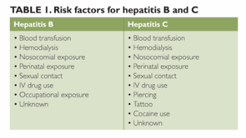
Infectious hepatitis is easily ruled in or out with serologic testing, which must take into consideration the patient's risk factors. The risk factors for hepatitis A, B, and C are presented in Table 1. Whether the patient has risk factors or not, these diseases should always be considered in such applicable populations as those with history of IV drug use (for hepatitis C virus [HCV] and hepatitis B virus [HBV]), men who have had sex with men (HBV), children born to a mother with HBV or HCV, and persons living in an area endemic for HBV.
ca0211liverfeat_t1_227422

Therapy for acute hepatitis comprises rest, nutrition, and fluids. A few cases of acute hepatitis will progress to acute liver failure. Prompt referral to a specialist is warranted for acute hepatitis that is not improving. Prevention of infectious hepatitis via vaccination is always recommended for appropriate individuals, such as health-care workers, people with liver disease or other chronic disease and those traveling to endemic areas.

Chronic hepatitis presents with mild AST/ALT elevations. According to the World Health Organization (WHO), approximately 350 million people live with chronic HBV infection3 and approximately 3% of the world's population has been infected with HCV.4

Hepatitis B. Patients who have chronic HBV infection may present with abnormal liver enzyme levels, fatigue, malaise, poor appetite and right upper-quadrant abdominal pain. Hepatitis B is diagnosed by serologic testing. The testing process is complex, and diagnosis relies on a number of assays, including those for hepatitis antigens (hepatitis B surface antigen [HBsAg] and hepatitis Be antigen [HBeAg]) and their respective antibodies (anti-HBsAg and anti-HBeAg), as well as antibodies to hepatitis B core antigen (anti-HBc immunoglobulin [Ig] M and IgG), and hepatitis B DNA.

The combination of results from all these assays will determine the diagnosis. For example, a patient who tests positive for HBsAg may have an acute or a chronic infection; results of the other assays will help to differentiate acute from chronic disease and determine the patient's immune status. Table 2 provides a summary of possible results and their interpretation.

ca0212liverfeat_t2_227426

Most adults clear HBV before it progresses to chronic infection. However, for the patient who develops chronic disease, several medical therapies are available, including both oral and subcutaneous injection treatments, such as pegylated interferon alpha 2a (Pegasys), entecavir (Baraclude), adefovir (Hespera), telbivudine (Tyzeka), and tenofovir (Viread). Therapy should be started when the ALT elevation is more than two times the upper limit of normal, the patient has decompensated cirrhosis, or liver biopsy shows evidence of disease.5,6 Treatment of chronic hepatitis B requires the guidance of a specialist.

Hepatitis C. The diagnosis of HCV infection is also based on serologic testing. Although several tests can be involved, the testing process is less complex than it is for HBV infection. Testing for HCV usually starts with an assay for the HCV antibody. This is only a screening test and is not diagnostic of HCV infection. The HCV recombinant strip immunoblot assay (RIBA) can help differentiate between exposure to HCV and actual infection.

Patients who test positive for HCV antibody need to be tested for virus in the serum. HCV RNA polymerase chain reaction (PCR) quantitative or HCV RNA qualitative testing is usually done next. Results of the qualitative test will be positive if any virus is detected in the blood, and the quantitative test will report the actual viral content. Some tests can detect a result as low as 43 IU/mL. A positive qualitative or quantitative test result is diagnostic for HCV infection. The actual viral count does not impact the severity of the disease. A patient with a viral load of 3 million copies/mL may not have more severe disease than the patient with a viral load of 1,000 copies/mL.

Therapy for chronic HCV infection is more complex than for other forms of hepatitis. Currently approved therapies include either daily doses of interferon alfacon-1 (Infergen) or weekly administration of pegylated interferon (Pegasys, Peg-interferon) with ribavirin (Copegus, Rebetol, RibaTab, Ribasphere). Two new adjunct therapies — the protease inhibitors telaprevir (Incivek) and boceprevir (Victrelis) — were approved last year. These therapies are used in conjunction with pegylated interferon and ribavirin in treating HCV infection. They are currently approved for use in those with hepatitis C disease.

Telaprevir and boceprevir can be used in a person with cirrhosis, but the individual must have well-compensated disease as evidenced by a Child-Turcotte-Pugh classification score of "A." The protease inhibitors are not currently approved for use in the post-transplant population. The new triple therapy had doubled the rates of sustained viral response (SVR). The SVRs of therapies range from 20% to 80%, depending on the degree of fibrosis and the genotype of the disease.

Patients with a confirmed diagnosis of HCV infection must be referred to a specialist for further evaluation. The specialist may choose to treat the patient or to perform liver biopsy. Because genotype 1 disease has the poorest rates of SVR, a liver biopsy is usually done to assess the extent of fibrosis.

Treatment is recommended in those with more advanced disease (stage 2+ fibrosis). Biopsy is not routinely done in patients with genotype 2 or 3 disease. Response to therapy is greatest in genotype 2 and 3 disease, and treatment is recommended in eligible patients regardless of disease severity.

Autoimmune hepatitis. This form of hepatitis primarily affects women. The age at diagnosis varies. Autoimmune hepatitis has links to other autoimmune diseases, such as thyroiditis, Sjögren syndrome, and diabetes mellitus. At presentation, the patient may have either mild or significant elevations of the aminotransferases. Autoimmune hepatitis is also ruled out or in with laboratory testing. The serum test for autoimmune hepatitis is the antinuclear antibody test. A liver biopsy is often done to further diagnose the disease as well as to stage the extent of inflammation and fibrosis.

Treatment of autoimmune hepatitis usually consists of high-dose prednisone (started at around 40 mg/day) and possibly another immunosuppressant medication, such as azathioprine (Azasan, Imuran). The prednisone can be slowly tapered in some patients. Management of this disease requires the supervision of a specialist.

NAFLD and NASH. Another common etiology of mild aminotransferase elevations is nonalcoholic fatty liver disease (NAFLD) or a more progressive form of obesity-related liver disease called nonalcoholic steatohepatitis (NASH). NAFLD is an equal-opportunity disease, affecting 20% of adults and 5% of children.7

NASH results from fat deposition in the liver (steatosis). Possible sequelae to this disorder include cirrhosis and HCC.2 We are still learning why patients progress from NAFLD to NASH. Elevations in fatty acids/triglycerides, obesity, and insulin resistance have all been associated with NASH. (See Table 3 for a more extensive list of associated conditions.) While some data have linked elevated triglycerides with NASH, the problem likely occurs when fatty acids accumulate because the fatty acid supply overwhelms triglyceride synthesis.2

ca0212liverfeat_t3_227430

NAFLD and NASH will likely not progress within the first few months of diagnosis. Many clinicians will monitor a patient after initial workup and make medical recommendations to lose weight or control diabetes mellitus (if applicable). Patients whose liver enzymes normalize after modifying their risk factors can be monitored conservatively and may avoid a liver biopsy. If the aminotransferase elevations persist, however, the patient requires referral to a specialist for a liver biopsy and further evaluation.

Ultrasonography and MRI have become more sensitive to fatty infiltration in the liver. The gold standard in diagnosing NAFLD and NASH is a liver biopsy. Liver biopsy carries risk factors and should be ordered only after other workup has been completed. Figure 1 is an algorithm for the evaluation of patients suspected of having NAFLD.
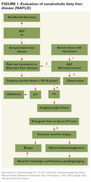
ca0212liverfeat_fig1_227434

Conclusion

Just as the liver has numerous functions, so does it have the potential for dysfunction. Primary-care clinicians with an understanding of basic liver function can begin the diagnostic process, recognizing that patients with more serious liver disorders may require the supervision of a hepatologist.

Rebecca Duke, MSN, APN, is a transplant nurse practitioner at Northwestern Medical Faculty Foundation in Chicago.

References

1. Bruix J, Sherman M. Management of hepatocellular carcinoma: an update. Hepatology. 2011;53:1020-1022.

2. Manns et al. American Association for the Study of Liver Diseases Diagnosis and Management of Autoimmune Hepatitis Guidelines. 2010.

3. World Health Organization. Hepatitis B.

4. Holmberg S. "Chapter 3: Infectious Diseases Related to Travel - Hepatitis C." CDC Yellow Book. 2012.

5. Lok AS, McMahon BJ. Chronic hepatitis B. Hepatology. 2001;34:1225-1241.

6. Lok AS, McMahon BJ. Chronic hepatitis B: update 2009. Hepatology. 2009;50:661-662.

7. Neuschwander-Tetri BA, Caldwell SH. Nonalcoholic steatohepatitis: summary of an AASLD Single Topic Conference. Hepatology. 2003;37:1202-1219.

Source